Monthly Archives: March 2017

MCL Works on Deep Learning based Fashion Fingerprinting

Fashion fingerprint is a compact feature vector for fashion items that can be used for tasks such as recognition, clustering, and retrieval of similar items. It is equally useful for both online fashion retailers as well as for physical apparel stores (with or without their online extensions). A related problem is understanding the apparel preferences of an individual from the dresses that they wear while visiting the physical store. One of the challenges of fashion study different from others is lack of enough accurate annotation. Available datasets have either limited number of images or very noisy annotation.

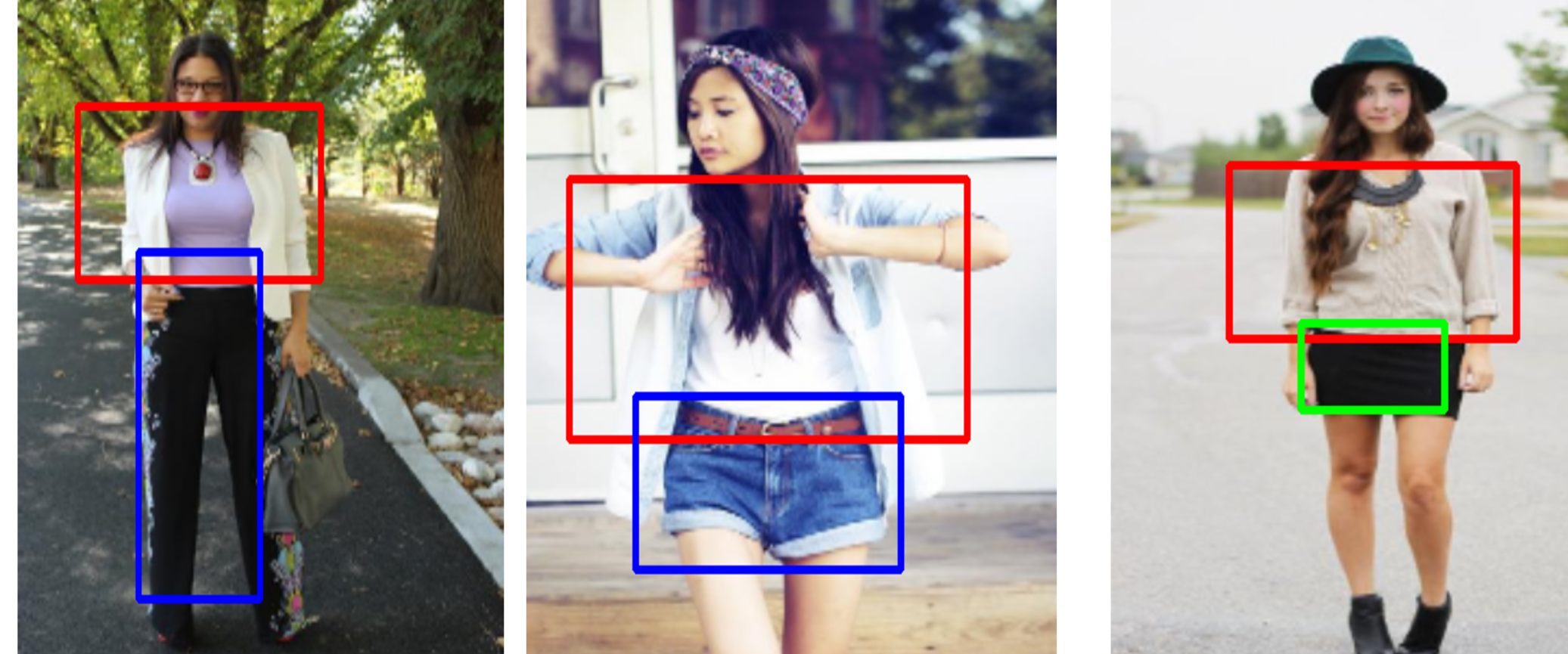
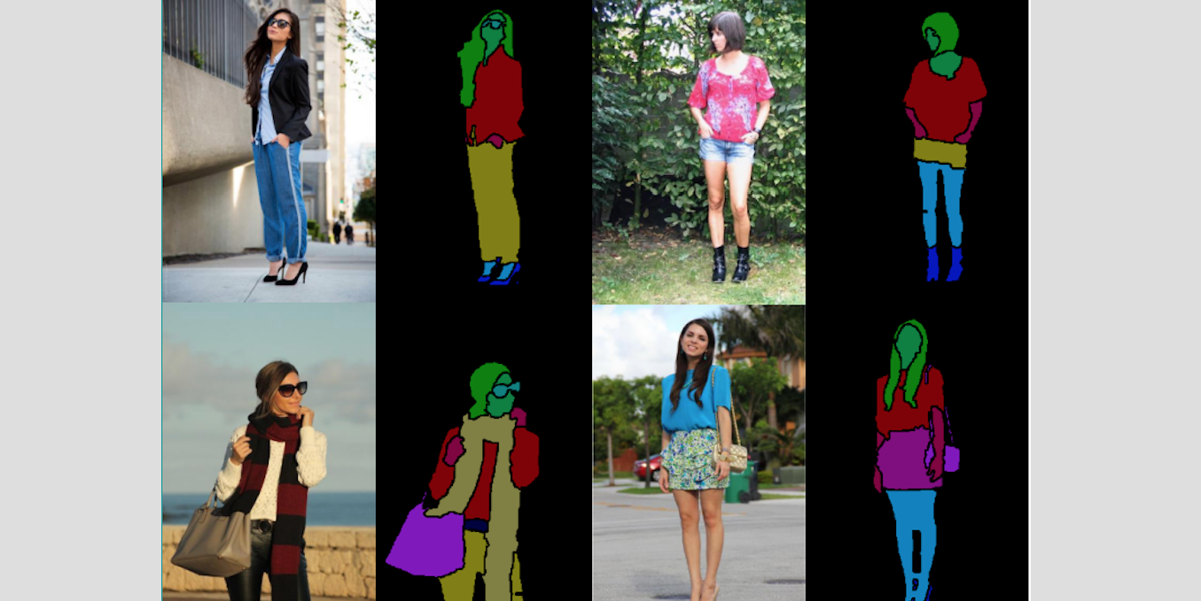
Currently we have successfully trained a fashion item localization model based on SSD[1]. The model is able to localize upper clothes, bottom clothes and one-pieces and has been tested on the Clothing Parsing dataset[2]. It achieves an F-score of 0.887 on upper clothes localization. For other clothing items, errors occur because the model may focus on too local regions and thus gets confused between skirt and dress. Prior location of human body will be incorporated in our model to solve this problem.

In the future we will further refine our localization model and also work on two directions. One is to recognize the garments based on our localization. The other one is to automatically label more images to enlarge the size of datasets.

[1] Liu, Wei, et al. “SSD: Single shot multibox detector.” European Conference on Computer Vision. Springer International Publishing, 2016.
[2] Liang, Xiaodan, et al. “Deep human parsing with active template regression.” IEEE transactions on pattern analysis and machine intelligence 37.12 (2015): 2402-2414.

By |March 26th, 2017|News|Comments Off on MCL Works on Deep Learning based Fashion Fingerprinting|

MCL Works on Splicing Image Detection

With the advent of Web 2.0 and ubiquitous adoption of low-cost and high-resolution digital cameras, users upload and share images on a daily basis. This trend of public image distribution and access to user-friendly editing software such as Photoshop and GIMP has made image forgery a serious issue. Splicing is one of the most common types of image forgery. It manipulates images by copying a region from one image (i.e., the donor image) and pasting it onto another image (i.e., the host or spliced image). Forgers often use splicing to give a false impression that there is an additional object present in the image, or to remove an object from the image. A spliced image from the Columbia Uncompressed [1] dataset is shown above. Image splicing can potentially be used in generating false propaganda for political purposes. For example, during the 2004 US Presidential election campaign, an image that showed John Kerry and Jane Fonda speaking together at an anti-Vietnam war protest was released and circulated. It was discovered later that this was a spliced image, and was created for political purposes. The spliced image and the two corresponding authentic images can be seen above [2].

Early work on image splicing detection only deduced whether a given image has been spliced or not, and no effort to localize the spliced area was attempted. The problem of joint splicing detection and localization has only been studied in recent years. For the problem of image splicing localization, one has to determine which pixels in an image have been manipulated as a result of a splicing operation.

One of the MCL members, Ronald Salloum, is currently working on an image splicing localization research project funded by the Defense Advanced [...]

By |March 22nd, 2017|News|Comments Off on MCL Works on Splicing Image Detection|
  • Permalink Gallery

    Congratulations to Professor Kuo for Receiving the 2017 IEEE Leon K. Kirchmayer Graduate Teaching Award

Congratulations to Professor Kuo for Receiving the 2017 IEEE Leon K. Kirchmayer Graduate Teaching Award

MCL Director, Professor C.-C. Jay Kuo, received the 2017 IEEE Leon K. Kirchmayer Graduate Teaching Award on March 6 at the 42nd IEEE International Conference on Acoustics, Speech and Signal Processing (ICASSP) held in New Orleans, Louisiana, USA.

The IEEE Leon K. Kirchmayer Graduate Teaching Award is sponsored by the Leon K. Kirchmayer Memorial Fund and recognizes inspirational teaching of graduate students in the IEEE fields of interest.  Professor Kuo received this award for excellence in inspirational guidance of graduate students and curriculum development in the area of multimedia signal processing.

Professor Kuo gave the following short speech in the Award Ceremony. “It was a great honor to be recognized by the prestigious Lion K. Kirchmayer Graduate Teaching Award. I would like to use this opportunity to appreciate a few people who had great impacts on my teaching career. When I was a PhD student at MIT, I was fortunate to work with a few young faculty members. They were my PhD and Master thesis advisor Bernard Levy, my PhD thesis co-advisor, John Tsitsiklis, my MS thesis co-advisor, Bruce Musicus, my PhD thesis committee member, Nick Trefethen, and my postdoc mentor at UCLA, Tony Chan. They spent an enormous amount of time nurturing and advising me. I am obliged to them deeply. Furthermore, I would like to say thanks to my graduate students. They are not only my students but also my teachers. I learned many new topics together with them. Finally, I would like to give thanks to my wife and daughter. Their unconditional love and patience allow me to do whatever I want to pursue. I do owe them tremendously, and would to like to share my joy and honor with them.”

Congratulations to Professor [...]

By |March 12th, 2017|News|Comments Off on Congratulations to Professor Kuo for Receiving the 2017 IEEE Leon K. Kirchmayer Graduate Teaching Award|
  • Permalink Gallery

    MCL Works on Automatic Medical Image Segmentation with Convolutional Neural Networks

MCL Works on Automatic Medical Image Segmentation with Convolutional Neural Networks

Automatic image segmentation has always been an important topic in medical imaging. Many medical applications, such as delineating heart structures, rely heavily on the accurate segmentation results. Nowadays, manual segmentation is still required in many applications. Manual segmentation is not only time-consuming and tedious but also prone to human error. One of MCL members, Ruiyuan Lin, is working on this research topic.

Many methods have been proposed to automate the segmentation process, ranging from region growing and active contour models to multi-atlas segmentation. In our research work, we focus on the convolutional neural networks (CNN) based segmentation method. We attempted several segmentation networks such as fully convolutional networks (FCN) and residual networks, compared their performance with other methods, and analyzed the strengths and problems of the networks. We are planning to further explore the use of CNN on more complicated medical images such as cross-domain images.

Image credit: both images are modified from the MRI images in the Left Atrium Segmentation Challenge dataset:
Tobon-Gomez C, Geers AJ, Peters, J, Weese J, Pinto K, Karim R, Ammar M, Daoudi A, Margeta J, Sandoval Z, Stender B, Zheng Y, Zuluaga, MA, Betancur J, Ayache N, Chikh MA, Dillenseger J-L, Kelm BM, Mahmoudi S, Ourselin S, Schlaefer A, Schaeffter T, Razavi R, Rhode KS. Benchmark for Algorithms Segmenting the Left Atrium From 3D CT and MRI Datasets. IEEE Transactions on Medical Imaging, 34(7):1460–1473, 2015.

By |March 5th, 2017|News|Comments Off on MCL Works on Automatic Medical Image Segmentation with Convolutional Neural Networks|