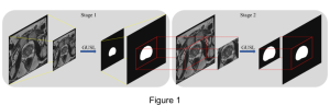
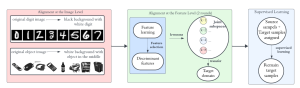
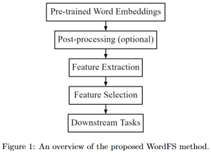
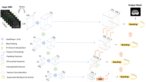
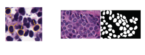
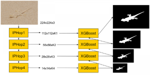
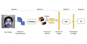
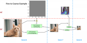
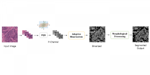
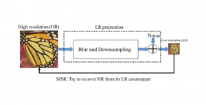
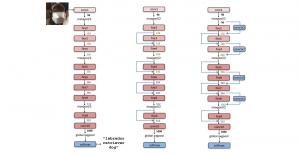
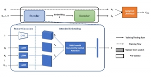
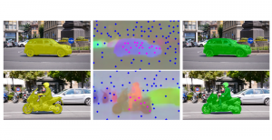
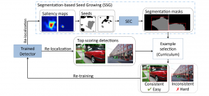
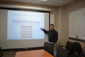
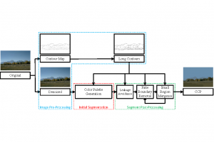
Automatic image segmentation has always been an important topic in medical imaging. Many medical applications, such as delineating heart structures, rely heavily on the accurate segmentation results. Nowadays, manual segmentation is still required in many applications. Manual segmentation is not only time-consuming and tedious but also prone to human error. One of MCL members, Ruiyuan Lin, is working on this research topic.
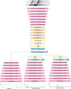
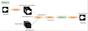
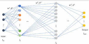
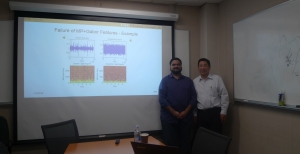
Many methods have been proposed to automate the segmentation process, ranging from region growing and active contour models to multi-atlas segmentation. In our research work, we focus on the convolutional neural networks (CNN) based segmentation method. We attempted several segmentation networks such as fully convolutional networks (FCN) and residual networks, compared their performance with other methods, and analyzed the strengths and problems of the networks. We are planning to further explore the use of CNN on more complicated medical images such as cross-domain images.
Image credit: both images are modified from the MRI images in the Left Atrium Segmentation Challenge dataset:
Tobon-Gomez C, Geers AJ, Peters, J, Weese J, Pinto K, Karim R, Ammar M, Daoudi A, Margeta J, Sandoval Z, Stender B, Zheng Y, Zuluaga, MA, Betancur J, Ayache N, Chikh MA, Dillenseger J-L, Kelm BM, Mahmoudi S, Ourselin S, Schlaefer A, Schaeffter T, Razavi R, Rhode KS. Benchmark for Algorithms Segmenting the Left Atrium From 3D CT and MRI Datasets. IEEE Transactions on Medical Imaging, 34(7):1460–1473, 2015.